Your gift is 100% tax deductible.
Thymus Cancer
If you have thymus cancer or are close to someone who does, knowing what to expect can help you cope. Here you can find out all about thymus cancer, including risk factors, symptoms, how it is found, and how it is treated.
About thymus cancer
Cancer that starts in the thymus gland is called thymus cancer. It starts when cells in the thymus gland grow out of control and crowd out normal cells. Thymus cancers are rare.
The thymus
The thymus is a small organ just behind the breastbone (sternum) in the front part of the chest. The thymus is in a part of the chest called the mediastinum. This is the space in the middle of the chest between the two lungs. The thymus sits just in front of and above the heart.
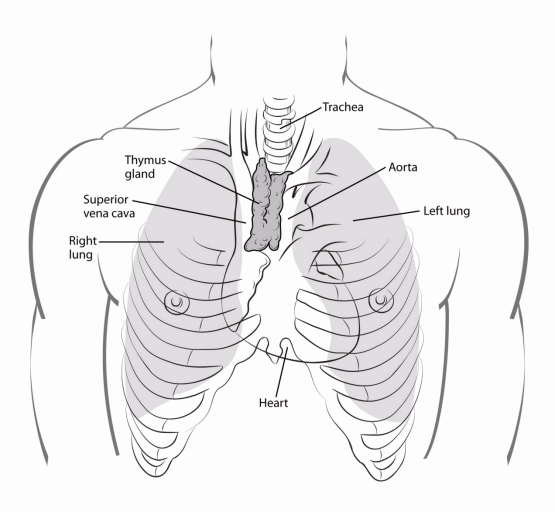
The thymus has 2 halves, called lobes. It has an irregular shape. There are a lot of small bumps called lobules on its surface. The thymus has 3 main layers:
- The medulla is the inner part of the thymus.
- The cortex is the outer layer that surrounds the medulla.
- The capsule is the thin covering over the outside of the thymus.
The thymus is largest during puberty. Then it shrinks during adulthood, and most of it is replaced by fat tissue. However, if the thymus stays large into adulthood, this does not always mean that cancer is present.
The thymus is an important part of the body’s lymphatic system. This is part of the immune system. Starting very early in life, the thymus makes immune cells called T lymphocytes and helps them to mature. You may hear these called T cells, named after the thymus. T lymphocytes then travel to lymph nodes (small, bean-shaped organs that filter lymph fluid) throughout the body. There they help fight infections.
The thymus is made of different types of cells. Each kind of cell can develop into a different type of cancer:
- Epithelial cells give the thymus its structure and shape. Thymomas and thymic carcinomas develop from these cells.
- Lymphocytes make up most of the rest of the thymus. Whether in the thymus or in other parts of the body, these immune system cells can develop into cancers called Hodgkin lymphoma and non-Hodgkin lymphoma.
- Kulchitsky cells, or neuroendocrine cells, are much less common cells that normally release certain hormones. These cells can develop into thymic neuroendocrine tumors (TNETs).
Thymomas and thymic carcinomas
Thymomas and thymic carcinomas are tumors that start from thymic epithelial cells. They are known as thymic epithelial tumors (TETs).
In the past, some thymomas were called benign (noncancerous). Doctors now consider all thymomas to be a form of cancer, even though they grow and spread slowly. What matters most is whether the cancer has grown beyond the thymus and how far it has grown. This is called the stage. Learn about the system used to describe the stage (extent) of thymomas in How Is Thymus Cancer Staged?
Thymic carcinomas tend to grow and spread faster than thymomas. They are grouped into subtypes, such as squamous cell carcinoma, based on how the cells look under a microscope.
Types of thymoma
Thymus cancers start in the cells that give the thymus its structure. They are classified by how they look under a microscope and by tests done on the tissue samples. Doctors call this the tumor type or histologic type. The World Health Organization (WHO) uses a lettered system to describe the different types.
Thymomas are divided into five main types.
Type AB is the most common type of thymoma. Type A is the least common.
The cells in these tumors can be spindle-shaped or oval epithelial cells. These cells are fairly normal looking. This is the rarest type of thymoma, but it seems to have the best outlook.
This type, also known as a mixed thymoma, looks mostly like type A. But there are also areas of lymphocyte cells mixed in the tumor.
This type looks a lot like the normal structure of the thymus. It has a lot of lymphocytes along with normal-looking thymus cells.
This type also has a lot of lymphocytes, but the thymus epithelial cells are larger, and the DNA-containing part of the cell (nuclei) looks abnormal.
This type has few lymphocytes and is mostly made of thymus epithelial cells that look close to normal.
Thymoma outlook
Types A, AB, and B1 tend to have a better outlook and usually grow slowly. Types B2 and B3 are more likely to grow quickly or spread to other parts of the body. Still, for most types of thymoma, the stage matters more than the type. The stage describes how the cancer has grown and spread.
Thymic carcinoma outlook
Thymic carcinoma cells look very abnormal under a microscope. The cells may no longer even look like thymus cells. Instead, the cells look like cancers that start in other parts of the body.
These tumors are often found after they have grown into nearby tissues or spread (metastasized) to other parts of the body. Because of this, the outlook for thymic carcinomas tends to be worse than for thymomas.
Thymic neuroendocrine tumors (TNETs)
Thymic neuroendocrine tumors (TNETs) are rare tumors that can form from neuroendocrine cells in the thymus. TNETs may be linked with other conditions that affect the endocrine system, like Cushing syndrome and MEN1 syndrome.
These tumors are rare, so not a lot is known about them. But they are usually treated like similar neuroendocrine tumors that start in other parts of the body.
There are several types of TNETs:
These are low-grade (slow growing) tumors that rarely spread.
These tumors tend to grow a little faster than typical carcinoids and are somewhat more likely to spread to other organs.
This is a high-grade cancer that tends to grow quickly and is likely to spread to other parts of the body.
This is a high-grade cancer, with cells that look like small cell cancers in other parts of the body, such as small cell lung cancer. These cancers tend to grow quickly and to spread to other parts of the body.
For more on how these tumors are typically treated, see Treating Lung Carcinoid Tumors and Treating Small Cell Lung Cancer.
Other cancers and tumors in the mediastinum
Other types of cancers and tumors can also grow in the mediastinum.
Cancers can start in the esophagus (esophageal cancer), in the heart (and the tissue surrounding it), in the trachea (windpipe), and in the lymph nodes (lymphoma).
Rarely, cancers and tumors known as germ cell tumors can start in the mediastinum. These come from cells like those found in the testicles and ovaries.
Sometimes the thyroid gland, which is normally in the neck, is misplaced into the mediastinum. If this happens, a thyroid tumor or cancer can develop in the mediastinum.
More often, cancer that starts in another part of the body, especially the lungs, spreads to the mediastinum.
- Written by
- References

Developed by the American Cancer Society medical and editorial content team with medical review and contribution by the American Society of Clinical Oncology (ASCO).
Detterbeck FC, Stratton K, Giroux D, et al. The IASLC/ITMIG Thymic Epithelial Tumors Staging Project: Proposal for an Evidence-Based Stage Classification System for the Forthcoming (8th) Edition of the TNM Classification of Malignant Tumors. J Thorac Oncol. 2014;9(9 Suppl 2):S65-72.
Filosso PL, Yao X, Ahmad U, et al. Outcome of Primary Neuroendocrine Tumors of the Thymus: A Joint Analysis of the International Thymic Malignancy Interest Group and the European Society of Thoracic Surgeons Databases. J Thorac Cardiovasc Surg. 2015;149(1):103-109.
Marx A, Chan JK, Chalabreysse L, et al. The 2021 WHO Classification of Tumors of the Thymus and Mediastinum: What Is New in Thymic Epithelial, Germ Cell, and Mesenchymal Tumors? J Thorac Oncol. 2022;17(2):200-213.
National Comprehensive Cancer Network. NCCN Clinical Practice Guidelines in Oncology (NCCN Guidelines): Thymomas and Thymic Carcinomas. Version 2.2026. Accessed at https://www.nccn.org on Jun 10, 2026.
Ozgun G, Nappi L. Primary Mediastinal Germ Cell Tumors: A Thorough Literature Review. Biomedicines. 2023;11(2):487.
Roden AC, Schoolmeester JK, Singh S, et al. Histologic Classification of Thymoma: A Practical Guide for Routine Cases. J Thorac Oncol. 2015;10(9):1259-1264.
Last Revised: August 12, 2026
American Cancer Society medical information is copyrighted material. For reprint requests, please see our Content Usage Policy.