
Your gift is 100% tax deductible.
Understanding Your Pathology Report
Biopsy samples are studied under the microscope by a pathologist, who is a specialist in diagnosing and classifying diseases. A pathology report gives a diagnosis for each sample taken and will be used to help manage your care.
What information is included in a pathology report?
After testing and analyzing your biopsy or cytology samples, the pathologist will send your cancer care team a report that gives a diagnosis for each sample. This report can be used to help manage your care.
The pathology report is often quite long and complex. It’s often divided into several sections.
The general identifying information includes:
- Your name
- Medical record number issued by the hospital
- Date when the biopsy or surgery was done
- Unique number of the sample, which is assigned in the lab
- Contact information for the pathologist, ordering provider, and the lab where the sample was tested
The next part of the report often contains information that was provided by the doctor who removed the tissue sample and/or the ordering provider. This may include:
- Your medical history
- Reason for the biopsy/surgery
- Any special requests that were made to the pathologist.
For example, if a lymph node sample was removed from a person with cancer in another organ, the doctor will note the original cancer type. The pathologist will order tests to learn if cancer in that lymph node is cancer that has spread or if it’s a new cancer that started in the lymph node.
The most important part of the pathology report is the final diagnosis. This section is usually at the top of the page. If the diagnosis is cancer, this section will note the exact type of cancer and will usually include the:
- Cancer’s grade and stage
- Lymph node status
- Margin status
Your cancer care team relies on this final diagnosis to help decide on the next steps of your care plan.
Sometimes, a biopsy result might be inconclusive, which means that the pathologist can’t provide a diagnosis based on the tests that were done. In such cases, the biopsy might need to be repeated, or other tests might be needed. If your biopsy result is inconclusive, talk to your cancer care team about what the next steps should be.
The pathologist provides a gross description by simply looking at, measuring, and feeling the tissue sample.
For a small biopsy, this description is often a few sentences noting the:
- Size
- Color
- Consistency
- Number of tissue-containing cassettes submitted for processing.
Larger biopsy or tissue samples will have much longer descriptions. For example, a mastectomy for breast cancer would include:
- Size of the entire sample
- Size of the tumor
- How many lymph nodes were found in the underarm area
- Appearance of the noncancerous tissue
- Summary of exactly where tissue was taken from.
When needed, the weight and number of samples are also included.
In some cases, multiple biopsies or tissue samples may be submitted and will be described individually, including how they were labeled and placed in cassettes.
For cytology samples, the gross description is very short and usually notes the number of slides or smears made by the doctor and how they were prepared. If the sample is a body fluid, its color and volume are noted.
This is a description of what the pathologist sees when looking at the processed sample under a microscope. It usually includes:
- Grade, or the appearance of the cancer cells
- Cell features, or how cancer cells are arranged or behave
- Invasion, or the extent to which the cancer invades nearby tissues
- Tumor margins, or if cancer cells are present at the edges of the sample
- Lymph node status, or if the lymph nodes were positive for cancer
Results of any other studies done, such as histochemical stains, flow cytometry, or biomarkers, may be noted in the microscopic description or in a separate section.
Some pathology reports may not have this section.
Pathology reports for cancers should contain a summary of the findings most relevant to making treatment decisions.
The College of American Pathologists (CAP) has guidelines that specify what information is most relevant for each type of cancer, and it provides templates of these “cancer protocols” to pathologists.
After the diagnosis, the pathologist may add more information for the doctor(s) taking care of the patient. The comment section is often used to clarify a concern or to recommend further testing.
This section may also include:
- Images
- References
- Other molecular studies
Sometimes, a pathologist is certain that a tissue sample is cancer based on the microscopic exam, but further testing is needed to determine the exact type of cancer or to provide the treating doctor with more information to help determine the best treatment.
If the additional testing is going to take several more days, the pathologist might issue an initial report stating the cancer diagnosis. Then, once further testing is complete, an addendum section is added to the report, which includes the results of these tests.
How to learn more or get a second opinion about your pathology results
Pathology results often play a key role when making decisions about treatment, and many people want to learn more about their test results.
Who should I ask about my pathology results?
Ask someone on your cancer care team to explain the results to you in a way that you can understand. Have them focus on how the results might impact your treatment options or your outlook.
Who has access to my pathology report?
All members of your cancer care team at the center where your cancer was diagnosed will have access to the pathology report and other medical records.
If you see doctors who practice at other facilities, such as for a second opinion, you might need to send copies of your pathology reports and other medical records before your appointment.
In most cases, you can sign a release form to have the copies sent, but it’s a good idea to keep an original copy for times you might need it again.
What if I need a second opinion from a pathologist?
If you or your cancer care team have any concerns about your diagnosis, you can have your samples reviewed by a consulting pathologist for a second opinion.
Your oncologist, surgeon, or the original pathologist can often suggest a consultant with special training in examining samples like yours. Or you can have your samples sent to the pathology department of a cancer center you have confidence in.
If your care is provided at an academic center, complicated cases may be reviewed in multidisciplinary tumor boards, where other pathologists, oncologists, and surgeons can review your case.
Some pathology labs will give you your processed samples, often on microscope slides, if you are going to visit another cancer center for a second opinion or consultation. Other labs will mail the microscope slides or send digital images directly to the consulting cancer center’s pathology department. You’ll probably have to sign forms to get this done.
Check with your cancer care team or the consulting doctor’s office on what you need to do.
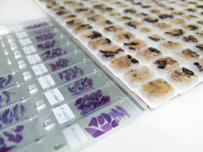
Stained microscope slides and biopsy tissue in paraffin blocks
- Written by
- References

Developed by the American Cancer Society medical and editorial content team with medical review and contribution by the American Society of Clinical Oncology (ASCO).
American Society of Clinical Oncology (ASCO). Reading a pathology report. Cancer.net. Content is no longer available.
College of American Pathologists. How to reach your pathology report. 2021. Accessed at https://documents.cap.org/documents/how-to-read-pathology-report.pdf on March 18, 2026.
National Cancer Institute (NCI). How cancer is diagnosed. Accessed from www.cancer.gov/about-cancer/diagnosis-staging/diagnosis on March19, 2026.
National Cancer Institute (NCI). Pathology reports. Updated August 8, 2022. Accessed at https://www.cancer.gov/about-cancer/diagnosis-staging/diagnosis/pathology-reports-fact-sheet#how-is-tissue-obtained-for-examination-by-a-pathologist on March 18, 2026.
Last Revised: March 26, 2026
American Cancer Society medical information is copyrighted material. For reprint requests, please see our Content Usage Policy.
American Cancer Society Emails
Sign up to stay up-to-date with news, valuable information, and ways to get involved with the American Cancer Society.

