
Your gift is 100% tax deductible.
Lung Neuroendocrine Tumor Stages
After someone is diagnosed with a lung neuroendocrine tumor (NET), doctors will try to figure out if it has spread, and if so, how far. This process is called staging.
The stage of a cancer describes how much cancer is in the body. It helps determine how serious the cancer is and how best to treat it. Doctors also use a cancer's stage when talking about survival statistics.
How is the stage determined?
The staging system most often used for lung NETs is the American Joint Committee on Cancer (AJCC) TNM system, which is based on 3 key pieces of information:
- The size and extent of the main tumor (T): How large is the tumor? Has it grown into nearby structures or organs?
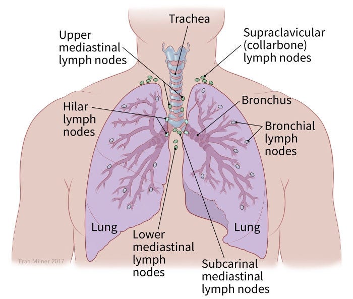
- The spread to nearby lymph nodes (N): Has the cancer spread to nearby lymph nodes?
- The spread (metastasis) to distant sites (M): Has the cancer spread to distant organs, like the liver?
Numbers or letters after T, N, and M provide more details about each of these factors. Higher numbers mean the cancer is more advanced. Once a person’s T, N, and M categories have been determined, this information is combined in a process called stage grouping to assign an overall stage. For more information, see Cancer Staging.
There is not a separate staging system for lung NETs. It uses the same classification as non-small cell lung cancer and small cell lung cancer.
Clinical vs. pathologic stage for lung neuroendocrine tumors
Lung NETs are typically given a clinical stage based on the results of physical exams, biopsies, imaging tests, and any other tests that have been done. If surgery is done, the pathologic stage, also called the surgical stage, is determined by examining tissue removed during the operation.
Stages of lung neuroendocrine tumors
The earliest stage is stage 0. The other main stages range from I (1) through IV (4). Some of these are divided further using letters or numbers. The lower the stage, the less the cancer has spread. Within a stage, an earlier letter or number means a lower stage. Although each person’s cancer experience is unique, cancers with similar stages tend to have a similar outlook and are often treated in much the same way.
Stage grouping: TX, N0, M0
The main tumor can’t be assessed for some reason. Or, cancer cells are seen in a sample of sputum or other lung fluids but the cancer isn’t found with other tests, so its location can’t be determined (TX). The cancer is not thought to have spread to nearby lymph nodes (N0) or to distant parts of the body (M0).
Stage grouping: Tis, N0, M0
The tumor is found only in the top layers of cells lining the air passages and has not invaded deeper into other lung tissues (Tis). The cancer has not spread to nearby lymph nodes (N0) or to distant parts of the body (M0).
Stage IA1 (stage grouping: T1a, N0, M0)
The tumor is no larger than 1 cm across. It has not reached the membranes that surround the lungs and it does not affect the main branches of the bronchi (T1a). The cancer has not spread to nearby lymph nodes (N0) or to distant parts of the body (M0).
Stage IA2 (T1b, N0, M0)
The tumor is larger than 1 cm but no larger than 2 cm across. It has not reached the membranes that surround the lungs and it does not affect the main branches of the bronchi (T1b). The cancer has not spread to nearby lymph nodes (N0) or to distant parts of the body (M0).
Stage IA3 (T1c, N0, M0)
The tumor is larger than 2 cm but no larger than 3 cm across. It has not reached the membranes that surround the lungs and it does not affect the main branches of the bronchi (T1c). The cancer has not spread to nearby lymph nodes (N0) or to distant parts of the body (M0).
Stage IB (T2a, N0, M0)
The tumor has one or more of the following features (T2a):
- It is larger than 3 cm but not larger than 4 cm across.
- It has grown into a main bronchus, but is not within 2 cm of the carina (the point where the windpipe splits into the left and right main bronchi) and it is not larger than 4 cm across.
- It has grown into the visceral pleura (the membranes surrounding the lungs) and is not larger than 4 cm across.
- It is partially clogging the airways (and is not larger than 4 cm across).
The cancer has not spread to nearby lymph nodes (N0) or to distant parts of the body (M0).
Stage IIA (stage grouping: T2b, N0, M0)
The tumor has one or more of the following features (T2b):
- It is larger than 4 cm but not larger than 5 cm across.
- It has grown into a main bronchus, but is not within 2 cm of the carina (the point where the windpipe splits into the left and right main bronchi) and it is larger than 4 cm but not larger than 5 cm across.
- The tumor has grown into the visceral pleura (the membranes surrounding the lungs) and is larger than 4 cm but not larger than 5 cm across.
- The tumor is partially clogging the airways and is larger than 4 cm but not larger than 5 cm across.
The cancer has not spread to nearby lymph nodes (N0) or to distant parts of the body (M0).
Stage IIB
T1a/T1b/T1c, N1, M0
The tumor is no larger than 3 cm across, has not grown into the membranes that surround the lungs, and does not affect the main branches of the bronchi (T1). It has spread to lymph nodes within the lung and/or around the area where the bronchus enters the lung (hilar lymph nodes). These lymph nodes are on the same side as the cancer (N1). The cancer has not spread to distant parts of the body (M0).
OR
T2a/T2b, N1, M0
The tumor has one or more of the following features (T2):
- It is larger than 3 cm but not larger than 5 cm across.
- It has grown into a main bronchus, but is not within 2 cm of the carina (the point where the windpipe splits into the left and right main bronchi) and it is not larger than 5 cm across).
- It has grown into the visceral pleura (the membranes surrounding the lungs) and is not larger than 5 cm.
- It is partially clogging the airways (and is not larger than 5 cm).
The cancer has also spread to lymph nodes within the lung and/or around the area where the bronchus enters the lung (hilar lymph nodes). These lymph nodes are on the same side as the cancer (N1). The cancer has not spread to distant parts of the body (M0).
OR
T3, N0, M0
The tumor has one or more of the following features (T3):
- It is larger than 5 cm but not larger than 7 cm across.
- It has grown into the chest wall, the inner lining of the chest wall (parietal pleura), the phrenic nerve, or membranes of the sac surrounding the heart (parietal pericardium).
- There are 2 or more separate tumor nodules in the same lobe of a lung.
The cancer has not spread to nearby lymph nodes (N0) or distant parts of the body (M0).
IIIA
Stage grouping: T1a/T1b/T1c, N2, M0
The cancer is no larger than 3 cm across, has not grown into the membranes that surround the lungs, and does not affect the main branches of the bronchi (T1). The cancer has spread to lymph nodes around the carina (the point where the windpipe splits into the left and right bronchi) or in the space between the lungs (mediastinum). These lymph nodes are on the same side as the main lung tumor (N2). The cancer has not spread to distant parts of the body (M0).
OR
T2a/T2b, N2, M0
The tumor has one or more of the following features (T2):
- It is larger than 3 cm but not larger than 5 cm across.
- It has grown into a main bronchus, but is not within 2 cm of the carina (the point where the windpipe splits into the left and right main bronchi) and it is not larger than 5 cm across).
- It has grown into the visceral pleura (the membranes surrounding the lungs) and is not larger than 5 cm.
- It is partially clogging the airways and is not larger than 5 cm.
The cancer has spread to lymph nodes around the carina (the point where the windpipe splits into the left and right bronchi) or in the space between the lungs (mediastinum). These lymph nodes are on the same side as the main lung tumor (N2). The cancer has not spread to distant parts of the body (M0).
OR
T3, N1, M0
The tumor has one or more of the following features (T3):
- It is larger than 5 cm but not larger than 7 cm across.
- It has grown into the chest wall, the inner lining of the chest wall (parietal pleura), the phrenic nerve, or membranes of the sac surrounding the heart (parietal pericardium).
- There are 2 or more separate tumor nodules in the same lobe of a lung.
The cancer has also spread to lymph nodes within the lung and/or around the area where the bronchus enters the lung (hilar lymph nodes). These lymph nodes are on the same side as the cancer (N1). The cancer has not spread to distant parts of the body (M0).
OR
T4, N0 or N1, M0
The tumor has one or more of the following features (T4):
- It is larger than 7 cm across.
- It has grown into the space between the lungs (mediastinum), the heart, the large blood vessels near the heart (such as the aorta), the windpipe (trachea), the tube connecting the throat to the stomach (esophagus), the thin muscle separating the chest from the abdomen (diaphragm), the backbone, or the carina.
- There are 2 or more separate tumor nodules in different lobes of the same lung.
The cancer may or may not have spread to lymph nodes within the lung and/or around the area where the bronchus enters the lung (hilar lymph nodes). Any affected lymph nodes are on the same side as the cancer (N0 or N1). The cancer has not spread to distant parts of the body (M0).
IIIB
T1a/T1b/T1c, N3, M0
The cancer is no larger than 3 cm across, has not grown into the membranes that surround the lungs, and does not affect the main branches of the bronchi (T1). The cancer has spread to lymph nodes near the collarbone on either side of the body, and/or has spread to hilar or mediastinal lymph nodes on the other side of the body from the main tumor (N3). The cancer has not spread to distant parts of the body (M0).
OR
T2a/T2b, N3, M0
The tumor has one or more of the following features (T2):
- It is larger than 3 cm but not larger than 5 cm across.
- It has grown into a main bronchus, but is not within 2 cm of the carina (the point where the windpipe splits into the left and right main bronchi) and it is not larger than 5 cm across.
- It has grown into the visceral pleura (the membranes surrounding the lungs) and is not larger than 5 cm.
- It is partially clogging the airways (and is not larger than 5 cm).
The cancer has spread to lymph nodes near the collarbone on either side of the body, and/or has spread to hilar or mediastinal lymph nodes on the other side of the body from the main tumor (N3). The cancer has not spread to distant parts of the body (M0).
OR
T3, N2, M0
The tumor has one or more of the following features (T3):
- It is larger than 5 cm but not larger than 7 cm across.
- It has grown into the chest wall, the inner lining of the chest wall (parietal pleura), the phrenic nerve, or membranes of the sac surrounding the heart (parietal pericardium).
- There are 2 or more separate tumor nodules in the same lobe of a lung.
The cancer has spread to lymph nodes around the carina (the point where the windpipe splits into the left and right bronchi) or in the space between the lungs (mediastinum). These lymph nodes are on the same side as the main lung tumor (N2). The cancer has not spread to distant parts of the body (M0).
T4, N2, M0
The tumor has one or more of the following features (T4):
- It is larger than 7 cm across.
- It has grown into the space between the lungs (mediastinum), the heart, the large blood vessels near the heart (such as the aorta), the windpipe (trachea), the tube connecting the throat to the stomach (esophagus), the thin muscle separating the chest from the abdomen (diaphragm), the backbone (spine), or the carina (the point where the windpipe splits into the left and right bronchi).
- There are 2 or more separate tumor nodules in different lobes of the same lung.
The cancer has spread to lymph nodes around the carina (the point where the windpipe splits into the left and right bronchi) or in the space between the lungs (mediastinum). These lymph nodes are on the same side as the main lung tumor (N2). The cancer has not spread to distant parts of the body (M0).
IIIC
T3, N3, M0
The tumor has one or more of the following features (T3):
- It is larger than 5 cm but not larger than 7 cm across.
- It has grown into the chest wall, the inner lining of the chest wall (parietal pleura), the phrenic nerve, or membranes of the sac surrounding the heart (parietal pericardium).
- There are 2 or more separate tumor nodules in the same lobe of a lung.
The cancer has spread to lymph nodes near the collarbone on either side of the body, and/or has spread to hilar or mediastinal lymph nodes on the other side of the body from the main tumor (N3). The cancer has not spread to distant parts of the body (M0).
T4, N3, M0
The tumor has one or more of the following features (T4):
- It is larger than 7 cm across.
- It has grown into the space between the lungs (mediastinum), the heart, the large blood vessels near the heart (such as the aorta), the windpipe (trachea), the tube connecting the throat to the stomach (esophagus), the thin muscle separating the chest from the abdomen (diaphragm), the backbone (spine), or the carina (the point where the windpipe splits into the left and right bronchi).
- There are 2 or more separate tumor nodules in different lobes of the same lung.
The cancer has spread to lymph nodes near the collarbone on either side of the body, and/or has spread to hilar or mediastinal lymph nodes on the other side of the body from the main tumor (N3). The cancer has not spread to distant parts of the body (M0).
IVA
Any T, Any N, M1a
The cancer can be any size and may or may not have grown into nearby structures (any T). It may or may not have reached nearby lymph nodes (any N). In addition, any of the following is true (M1a):
- The cancer has spread to the other lung.
- Cancer cells are found in the fluid around the lung (called a malignant pleural effusion).
Cancer cells are found in the fluid around the heart (called a malignant pericardial effusion).
OR
Any T, Any N, M1b
The cancer can be any size and may or may not have grown into nearby structures (any T). It may or may not have reached nearby lymph nodes (any N). It has spread as a single tumor outside of the chest, such as to a distant lymph node or an organ such as the liver, bones, or brain (M1b).
IVB
Any T, Any N, M1c
The cancer can be any size and may or may not have grown into nearby structures (any T). It may or may not have reached nearby lymph nodes (any N). It has spread as more than one tumor outside the chest, such as to distant lymph nodes and/or to other organs such as the liver, bones, or brain (M1c).
*The following additional categories are not listed in the table above:
- T0: There is no evidence of a primary tumor.
- NX: Nearby lymph nodes cannot be assessed due to lack of information.
- Written by
- References

Developed by the American Cancer Society medical and editorial content team with medical review and contribution by the American Society of Clinical Oncology (ASCO).
American Joint Committee on Cancer. Lung. In: AJCC Cancer Staging System. Version Nine. American College of Surgeons; 2024.
Last Revised: December 17, 2025
American Cancer Society medical information is copyrighted material. For reprint requests, please see our Content Usage Policy.
American Cancer Society Emails
Sign up to stay up-to-date with news, valuable information, and ways to get involved with the American Cancer Society.

