
Your gift is 100% tax deductible.
Gallbladder Cancer Stages
After a person is diagnosed with gallbladder cancer, doctors will try to figure out if it has spread, and if so, how far. This process is called staging. The stage of a cancer describes how much cancer is in the body. It helps determine how serious the cancer is and how best to treat it. Although each person’s cancer experience is unique, cancers with similar stages tend to have a similar outlook and are often treated in much the same way. Doctors also use a cancer's stage when talking about survival statistics.
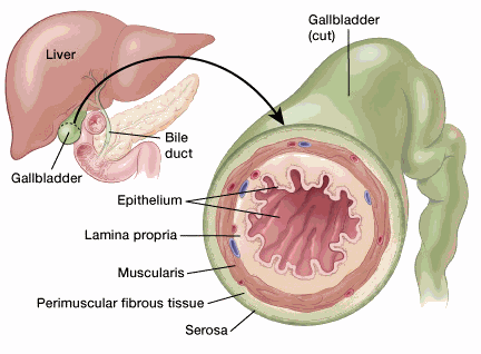
Nearly all gallbladder cancers start in the epithelium (the inside wall of the gallbladder). Over time, they grow through the various layers toward the outside of the gallbladder. They may also grow to fill up some or all the space inside the gallbladder at the same time. The gallbladder wall has several layers. From the inside out, these are:
- The epithelium, a thin sheet of cells that line the inside wall of the gallbladder
- The lamina propria, a thin layer of loose connective tissue (the epithelium plus the lamina propria form the mucosa)
- The muscularis propria, a layer of muscular tissue that helps the gallbladder contract to push its bile into the bile duct
- The perimuscular connective tissue, a layer of connective tissue around the muscle
- The serosa, the outer covering of the gallbladder that comes from the peritoneum, which is the lining of the abdominal cavity
The depth that a tumor grows from the inside (epithelium layer) through the other outer layers (all the way through the serosa) is a key part of staging.
How to read AJCC gallbladder cancer stage groupings?
The staging system most often used for gallbladder cancer is the American Joint Committee on Cancer (AJCC) TNM system, which is based on 3 key pieces of information:
- The extent (size) of the tumor (T): How far has the cancer grown into the wall of the gallbladder? Has the cancer grown through the gallbladder wall into nearby organs such as the liver?
- The spread to nearby lymph nodes (N): Has the cancer spread to nearby lymph nodes, and if so, how many?
- The spread (metastasis) to distant sites (M): Has the cancer spread to distant organs such as the liver, the peritoneum, or the lungs?
The gallbladder staging system uses the pathologic stage (also called the surgical stage), which is determined by tissue removed during an operation. Sometimes, if surgery can't be done right away or at all, the cancer will be given a clinical stage instead. This is based on the results of a physical exam, biopsy, and imaging tests. The clinical stage will be used to help plan treatment. Sometimes, the cancer has spread further than the clinical stage estimates, and it may not predict the patient’s outlook as accurately as a pathologic stage.
The system described below is the most recent AJCC system, effective January 2018. This system is used to stage cancers of the gallbladder and cancers that start in the cystic duct (the tube that carries bile away from the gallbladder).
The earliest stage gallbladder cancers (called carcinoma in situ) are stage 0. Stages then range from stages I (1) through IV (4). As a rule, the lower the number, the less the cancer has spread. A higher number, such as stage IV, means the cancer has spread more. And within a stage, an earlier letter means a lower stage.
Numbers or letters after T, N, and M provide more details about each of these factors. Higher numbers mean the cancer is more advanced.
Once a person’s T, N, and M categories have been determined, this information is combined in a process called stage grouping to assign an overall stage. For more on this, see Cancer Staging.
Cancer staging can be complex, so ask your doctor to explain it to you in a way you understand.
AJCC Stage |
Stage grouping |
Stage description* |
0 |
Tis N0 M0 |
Cancer is only in the epithelium (the inner layer of the gallbladder) and has not grown into deeper layers of the gallbladder (Tis). It has not spread to nearby lymph nodes (N0) or to distant parts of the body (M0). |
I |
T1 N0 M0 |
The tumor has grown into the lamina propria or the muscle layer (muscularis) (T1). It has not spread to nearby lymph nodes (N0) or to distant parts of the body (M0). |
IIA |
T2a N0 M0 |
The cancer has grown through the muscle layer into the fibrous tissue on the side of the peritoneum (the lining of the abdominal cavity) (T2a). It has not spread to nearby lymph nodes (N0) or to distant parts of the body. (M0). |
IIB |
T2b N0 M0 |
The cancer has grown through the muscle layer into the fibrous tissue on the side of the liver, but has not invaded the liver (T2b). It has not spread to nearby lymph nodes (N0) or to distant parts of the body (M0). |
IIIA |
T3 N0 M0 |
The cancer has grown through the serosa (the outermost covering of the gallbladder) and/or it has grown directly into the liver and/or one nearby structure like the stomach, duodenum (first part of the small intestine), colon, pancreas, or bile ducts outside the liver (T3). It has not spread to nearby lymph nodes (N0) or to distant parts of the body (M0). |
IIIB |
T1-3 N1 M0 |
The cancer may or may not have grown outside of the gallbladder into the liver and/or one other nearby structure, but it has not grown into the main blood vessels leading into the liver (portal vein or hepatic artery) (T1 to T3). It has spread to no more than 3 nearby lymph nodes (N1). It has not spread to distant parts of the body (M0). |
IVA |
T4 N0 or N1 M0 |
The tumor has grown into one of the main blood vessels leading into the liver (portal vein or hepatic artery) or it has grown into 2 or more structures outside of the liver (T4). It may or may not have spread to no more than 3 nearby lymph nodes (N0 or N1). It has not spread to distant parts of the body (M0). |
IVB |
Any T N2 M0 |
The primary tumor may or may not have grown outside the gallbladder. The cancer has spread to 4 or more nearby lymph nodes (N2). It has not spread to distant parts of the body (M0). |
OR |
||
Any T Any N M1 |
The primary tumor may or may not have grown outside the gallbladder. The cancer may or may not have spread to nearby lymph nodes. It has spread to distant places such as the liver, peritoneum (the lining of the abdomen [belly]), or the lungs (M1). |
|
* The following additional categories are not listed on the table above:
- TX: Main tumor cannot be assessed due to lack of information.
- T0: No sign of a primary tumor.
- NX: Regional lymph nodes cannot be assessed due to lack of information.
Other prognostic factors
Besides your stage, there are other factors that can affect your prognosis (outlook).
Grade
The grade describes how closely the cancer cells look like normal gallbladder cells when seen with a microscope.
The scale used for grading gallbladder cancer is from 1 to 3.
- Grade 1 (G1) cancer cells look a lot like normal gallbladder cells.
- Grade 3 (G3) cancer cells look very abnormal.
- Grade 2 (G2) cancer cells fall somewhere in between.
Low-grade cancers (G1) tend to grow and spread more slowly than high-grade (G3) cancers. Most of the time, the outlook is better for Grade 1 and Grade 2 cancers than it is for Grade 3 cancers of the same stage for gallbladder cancer.
Subtype
The type of gallbladder cancer you have can influence your outlook. Rare cancer types such as squamous and adenosquamous carcinomas of the gallbladder tend to have a worse prognosis (outlook) than adenocarcinomas (the most common type) and papillary carcinomas.
Lymphovascular invasion
If cancer cells are seen in small blood vessels (vascular) or lymph vessels (lymphatics) under the microscope, it's called lymphovascular invasion. When cancer is growing in these vessels, there's a greater chance that it has spread outside the gallbladder. Gallbladder cancers with lymphovascular invasion tend to have a poor prognosis.
Extent of resection
Cancers that can be removed completely by surgery tend to have a better outlook than those that cannot.
- Resectable cancers are those doctors believe can be removed completely by surgery.
- Unresectable cancers have spread too far or are in too difficult a place to be removed entirely by surgery.
Only a small percentage of gallbladder cancers are resectable when they're first found.
- Written by
- References

Developed by the American Cancer Society medical and editorial content team with medical review and contribution by the American Society of Clinical Oncology (ASCO).
National Comprehensive Cancer Network. NCCN Clinical Practice Guidelines in Oncology: Biliary Tract Cancers. v.1.2025 - March 20, 2025. Accessed at https://www.nccn.org/professionals/physician_gls/pdf/btc.pdf on April 17, 2025.
Last Revised: May 16, 2025
American Cancer Society medical information is copyrighted material. For reprint requests, please see our Content Usage Policy.
American Cancer Society Emails
Sign up to stay up-to-date with news, valuable information, and ways to get involved with the American Cancer Society.

