
Your gift is 100% tax deductible.
How Is a Biopsy Done?
A biopsy can be done in several ways, depending on the area being tested and what type of sample is needed.
What are the types of biopsy tests and how are they done?
There are multiple ways that biopsy tests can be done. Depending on the type of test, different types of sedation and anesthesia might be used.
For details about biopsy tests for a specific cancer type, go to its webpage and see its Early Detection, Diagnosis, and Staging section.
Needle biopsy
In a needle biopsy, a hollow needle is used to remove small samples of tissue or fluid. There are 2 main types of needle biopsies:
FNA uses a very thin, hollow needle attached to a syringe to withdraw (aspirate) a small amount of tissue, and sometimes fluid, from the mass. This includes areas like lymph nodes, breast lumps, or thyroid nodules.
If the mass is near the skin and can be felt, the doctor can insert the needle directly into the area. If the mass is deeper inside the body, the needle can be guided into place using an imaging test such as an ultrasound or CT scan.
FNAs usually don’t cause much pain, but if the area to be sampled is sensitive, the doctor may inject local anesthesia (numbing medicine) under the skin before it is done.
The main advantages of FNA are that the skin doesn’t have to be cut, and in some cases, it’s possible to make a diagnosis the same day. The main drawback is that sometimes the thin needle can’t remove enough tissue to make a definite diagnosis or to do other lab tests that might be needed.
Although FNA is a type of biopsy, it can also be considered a type of cytology test.
CNBs use needles that take a small cylinder, or “core,” of tissue. An advantage of this type of biopsy is that it removes more tissue than an FNA without needing surgery.
A CNB is most often done using local anesthesia (numbing medicine) in a doctor’s office or clinic. It can be used to sample masses that the doctor can feel, as well as some that can only be seen using imaging tests.
Doctors sometimes use special vacuum tools to get larger core biopsies. They can collect multiple or larger samples from the same biopsy site with this method. This is called vacuum-assisted biopsy and is used in some types of breast biopsies.
Surgical (open) biopsy
Open biopsies use some type of surgery to remove samples for testing.
For these biopsies, a surgeon cuts through the skin and uses a scalpel to remove either:
- The entire mass, called an excisional biopsy.
- A small part of a large mass, called an incisional biopsy.
Choosing between an excisional or incisional biopsy depends on the mass’s size and location, the suspected type of cancer, and whether the tissue is being removed for treatment or to make a diagnosis. If a larger sample is needed, an excisional biopsy might be best.
These biopsies are often done as outpatient procedures, where you don’t have to stay overnight. Depending on the type of biopsy, they may be done in a same-day surgery center, hospital, or doctor’s office.
This is often done using local or regional anesthesia (medicines that numb the area). For masses that are deeper in the body, such as inside the chest or abdomen, you might be under general anesthesia (in a deep sleep). Once the sample is removed, the cut is stitched or glued shut.
A shave biopsy removes the top layers of skin by using a razor-like tool to scrape the surface. They are most often used for suspected basal cell or squamous cell skin cancers, and less often for suspected melanomas of the skin.
Punch biopsies use a small, circular tool that looks like a cookie cutter to push through the surface of the skin to take a sample of tissue from below the skin’s surface. This lets doctors learn how deeply a cancer, such as melanoma, has grown into the skin. After the tissue sample is removed, the edges of the site are closed using stitches.
Both types use local anesthesia (numbing medicine) near the area being removed and are often performed in doctor’s offices.
Learn more about different types of skin biopsies.
A laparotomy is a type of surgery in which the doctor cuts into the abdomen (belly), usually with a vertical cut from upper to lower abdomen. A thoracotomy is a similar procedure in which a long cut is made to look inside the chest.
These procedures might be done:
- When a suspicious area can’t be diagnosed with less invasive tests
- If the surgeon plans to do other things as well, such as removing a large mass, tumor, or organ.
During these procedures, biopsy samples can be taken from a suspicious area. The doctor can also look at the size of the area and its location, as well as check nearby tissues. Once the tissue sample is removed, the cut is closed using stitches.
This type of procedure typically requires you to be under general anesthesia (in a deep sleep).
Endoscopic biopsy
An endoscopic biopsy is done with a procedure known as an endoscopy.
During an endoscopy, a doctor uses an endoscope (a thin tube with a light and camera on the end) to look inside a person’s body. During an endoscopic biopsy, the doctor passes special tools down the endoscope to remove tissue or fluid samples for testing.
Sometimes the endoscope is passed through an opening of the body, such as the mouth, to look at a specific area. These can be done in the doctor’s office or clinic with or without sedation to help you relax, or less often, be under general anesthesia (in a deep sleep).
Other types of endoscopic biopsies can be done by passing the endoscope into the body through a small cut made in the skin. During these procedures, special tools can be passed through other small cuts to remove biopsy samples for testing.
These are most often done in an operating room using different types of anesthesia, depending on the type of biopsy being done.
Bone marrow biopsy and aspiration
A bone marrow aspiration takes a small sample of the liquid bone marrow from inside your bones, while a bone marrow biopsy takes a piece of bone and marrow.
Liquid biopsy
For a liquid biopsy, a sample of blood or another body fluid, such as the pleural fluid in the chest, is removed and tested for cancer. Some cancers shed tumor cells or parts of cells into the blood, which can be detected with special lab tests. Depending on the test, it might look for:
- Cancer cells, sometimes called circulating tumor cells, or CTCs
- Genetic material from the cells, such as RNA or DNA, sometimes called circulating tumor DNA (ctDNA) or cell-free DNA (cfDNA)
- Proteins from the cancer cells
Not all cancers shed cells or parts of cells into the blood, so these tests can’t always be used. But when a liquid biopsy can be done, it can often provide some of the same important information that a tissue biopsy can. Liquid biopsies might help:
- Find cancer early.
- Look for certain gene or protein changes in the cancer cells, which might help determine the best treatment options. This is an example of personalized or precision medicine.
- Determine how well treatment is working or if further treatment might be needed.
- Look for changes in the cancer cells over time, which may help determine if treatment needs to be changed.
- Look for signs that cancer has come back.
Doctors are still learning the best ways to use liquid biopsies. But these tests are becoming more important in treating some types of cancer.
Sentinel lymph node mapping and biopsy
Sentinel lymph node mapping and biopsy (SLNB) can find the lymph nodes that drain lymph fluid from the area where the cancer started, called sentinel lymph nodes. If the cancer has spread, it will likely go to these lymph nodes first. This can help determine the stage (extent) of the cancer and help guide treatment options.
To find (map) the sentinel lymph node(s), the doctor injects a small amount of radioactive material into the area of the cancer, sometimes along with a colored dye. The sentinel lymph node(s), which take up the radioactivity first, are located by a special machine. The doctor will then make a small cut in the skin to access and remove them to be looked at under a microscope.
If no cancer cells are found, no more lymph nodes need to be taken out because it’s very unlikely the cancer would have spread beyond this point. If cancer cells are found in the sentinel node, additional nearby lymph nodes in this area are usually removed and looked at too. This is called a lymph node dissection.
How are biopsy samples processed?
After a biopsy sample is removed, it must be looked at by a pathologist in the lab. Depending on the testing method, it may be sent fresh or in a liquid preservative, such as formaldehyde (formalin).
Gross examination
A gross examination is where a pathologist looks at the specimen without a microscope. During the gross examination, the tissue sample’s size, color, consistency, and other characteristics are recorded. Photos of the sample may also be taken as part of the record.
The gross examination is important because the pathologist may see features that suggest a particular diagnosis, such as cancer. It also helps the pathologist decide which parts of a large biopsy sample are the most critical to look at under a microscope.
For smaller types of biopsies, such as a punch biopsy or a core needle biopsy, the entire specimen is usually looked at under a microscope.
Processing the specimen for microscopic examination
The biopsy tissue is put into small containers called cassettes, which hold the tissue securely. Then, usually overnight, steps are taken to preserve and dehydrate the sample. After processing, the tissue sample is put into a mold with hot paraffin wax. The wax cools to form a solid block that protects the tissue.
This paraffin wax block with the embedded tissue is then cut into very thin slices, which are placed on glass slides. The slides are dipped into a series of stains or dyes to change the color of the tissue. The color makes different parts of the cells easier to see under a microscope.
At this point, the pathologist looks at the tissue under a microscope. Looking at the solid specimens in this way is called histology, which is the study of the structures of cells and tissues.
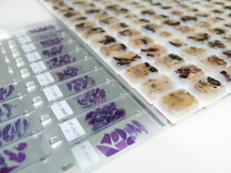
Stained histology slides and biopsy tissue in paraffin blocks
Frozen section (intra-operative consultation)
Sometimes during surgery, the surgeon will ask for an intra-operative (during surgery) pathology consult. This is often called a frozen section exam.
Why might a frozen section be done?
Frozen sections are typically done during surgery, when the surgeon needs information right away that can help guide what type of operation should be done. A frozen section might be done to:
Find out if a mass is cancer
Sometimes the type of surgery depends on whether the mass is cancer (malignant). For instance, if a frozen section shows the mass is not cancer (benign), removing the mass alone may be enough. But if it is cancer, the surgeon might need to remove a wider margin of tissue around the mass, as well as nearby lymph nodes. A frozen section can help guide this decision during surgery.
In some cases, the frozen section doesn’t give a clear answer, and the tissue needs more detailed testing. If this happens, the surgeon usually stops the operation and closes the cut. Once the results are back, another surgery may be needed.
Help ensure all the cancer has been removed
If the surgeon is concerned that a cancer hasn’t been removed completely, a slice from the margin (edge) of the tissue that was removed can be sent for a frozen section diagnosis:
- Negative margins mean that no cancer cells are seen at the margin and typically no more tissue needs to be removed. This doesn’t guarantee that the cancer won’t come back, although it makes it less likely.
- Positive margins mean that cancer cells are found at the margin, and it’s likely that at least some cancer cells were not removed.
If the margins are positive, the surgeon will usually remove more tissue from the area to try to get all the cancer cells and lower the chances of the cancer coming back.
Taking more tissue can help treat the cancer, but it also carries risks. The surgeon considers the benefits and possible complications. If it’s not possible to remove more tissue, there may be other treatment options, such as using radiation to kill the remaining cancer cells.
A frozen section can also be used as part of some surgical techniques, such as Mohs surgery (also called Mohs micrographic surgery, or MMS), for some skin cancers.
How is a frozen section done?
When a frozen section exam is done, fresh biopsy tissue is sent from the operating room directly to the pathologist. Because the patient is often still under general anesthesia, it’s important that the tissue is looked at quickly – usually within 10 to 20 minutes.
The pathologist will do a gross examination of the fresh tissue to decide which part of it should be looked at under a microscope. Instead of processing the tissue in wax blocks, the tissue is quickly frozen in a special solution that forms what looks like an ice cube around the tissue. It’s then sliced into very thin sections, quickly stained by dipping it in a series of dyes, and then looked at under a microscope.
The frozen sections typically don’t show features of the tissue quite as well as sections of tissue embedded in wax, but they are usually good enough to help the surgeon make decisions during the operation.
How long are pathology specimens kept?
Processed samples are usually kept for a set amount of time, which allows them to be sent to another doctor or lab for a second opinion. This process, called a pathology review, involves another doctor examining the sample to confirm or suggest a different diagnosis.
Why keeping specimens can be helpful
Keeping samples can also help if a new cancer occurs years later. Original and new slides can be compared to help determine if the cancer has come back (recurrence) or if it is new. Additional tests, like special stains on the original cells, might also be helpful.
Legal requirements
Clinical labs are regulated and certified based on a federal law called CLIA (Clinical Laboratory Improvement Amendments). To be CLIA accredited, labs must keep biopsy samples for a minimum amount of time. For instance, CLIA says that labs must keep:
- Histopathology slides for at least 10 years
- Paraffin blocks for at least 2 years
Some states have their own laws that require labs to keep biopsy samples longer than the time specified by CLIA. And some labs have policies for keeping samples even longer than required by federal or state laws.
If you would like to know if the lab you go to is CLIA certified, you can ask the lab staff. Or if you have questions or concerns, you can reach out to your state’s CLIA agency.
- Written by
- References

Developed by the American Cancer Society medical and editorial content team with medical review and contribution by the American Society of Clinical Oncology (ASCO).
American Society of Clinical Oncology. Biopsy. Accessed from www.cancer.net. Content no longer available.
Centers for Medicare & Medicaid Services. Clinical laboratory improvement amendments (CLIA). Cms.org. Updated February 6, 2026. Accessed at https://www.cms.gov/medicare/quality/clinical-laboratory-improvement-amendments on March 5, 2026.
National Cancer Institute (NCI). How cancer is diagnosed. Updated January 17, 2023. Accessed at https://www.cancer.gov/about-cancer/diagnosis-staging/diagnosis#biopsy on March 5, 2026.
National Institutes of Health (NIH). Biopsy. Updated November 18, 2025. Accessed at medlineplus.gov/biopsy on March 5, 2026.
Radiological Society of North America. General biopsy. Updated April 2024. Accessed at https://www.radiologyinfo.org/en/info/biopgen on March 5, 2026.
Slaoui M, Bauchet A-L, Fiette L. Tissue sampling and processing for histopathology evaluation. Methods Mol Bio. 2017; 1641:101-114.
Last Revised: March 24, 2026
American Cancer Society medical information is copyrighted material. For reprint requests, please see our Content Usage Policy.
American Cancer Society Emails
Sign up to stay up-to-date with news, valuable information, and ways to get involved with the American Cancer Society.

