Your gift is 100% tax deductible.
Core Needle Biopsy of the Breast
If exams or imaging tests show you might have breast cancer, your doctor might refer you for a core needle biopsy (CNB) to help find out for sure.
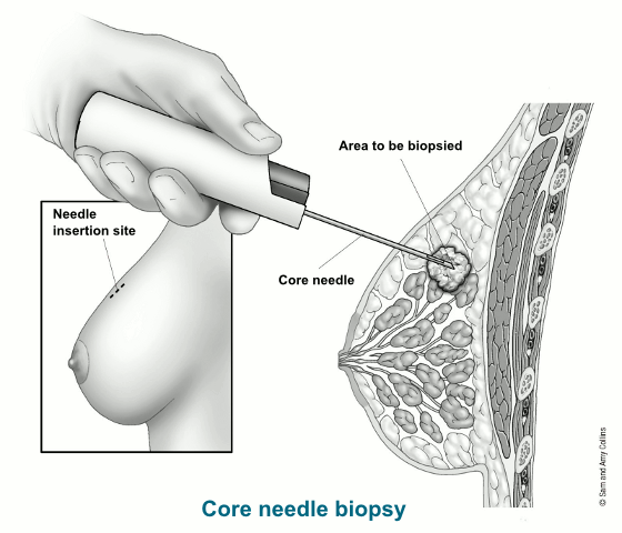
During a CNB, the doctor uses a hollow needle to take out pieces of breast tissue from the area of concern. The needle can be guided into the abnormal area by touch, if a lump can be felt, or by using an imaging test.
What is a core needle biopsy?
For a CNB, the doctor uses a hollow needle to take out pieces of breast tissue from a suspicious area the doctor has felt or seen on an imaging test. This is often the preferred type of biopsy if breast cancer is suspected, because it removes more breast tissue than a fine needle aspiration (FNA), but it does not require surgery.
The needle might be attached to a spring-loaded tool that moves the needle in and out of the tissue quickly, or it might be attached to a suction device that helps pull breast tissue into the needle, known as a vacuum-assisted core biopsy.
Typically, a tiny tissue marker, also called a clip, is put into the area where the biopsy is done. This marker will show up on mammograms or other imaging tests so the exact area can be located for further treatment or follow-up if needed. You can’t feel or see the marker. It stays in place, is safe during MRIs, and it will not set off metal detectors.
A small cylinder (core) of tissue is taken out in the needle. Several cores are often removed.
Getting the results
A doctor called a pathologist will look at the sample under the microscope to check whether there are cancer cells. A CNB is likely to clearly show whether cancer is present and often provides enough of a sample if other lab tests are needed, but it can still miss some cancers.
Ask your doctor when you can expect to get the results of your biopsy. If the results of the CNB do not give a clear diagnosis, or if your doctor still has concerns, you might need to have a second CNB or a more extensive type of biopsy, such as a surgical (open) biopsy.
Types of image-guided core needle biopsies
The doctor might put the needle into the abnormal area by feeling the lump. But usually some type of imaging test is used to guide the needle into the right place. Some of the imaging tests a doctor might use include:
- Mammogram or breast tomosynthesis, known as a stereotactic biopsy
- Ultrasound
- MRI
The type of image-guided biopsy that is best for you will depend on which type of imaging test will show the abnormal area best, as well as your comfort.
For this procedure, a doctor uses mammogram pictures taken from different angles to pinpoint the biopsy site. A computer analyzes the pictures and shows where the needle tip needs to go in the abnormal area. This type of biopsy is often used to check suspicious microcalcifications (tiny calcium deposits), small masses, or other abnormal areas that can’t be seen clearly on an ultrasound.
What to expect
You might be sitting up, lying on your side, or lying on your belly with your breast hanging through a hole in the table for this procedure.
The breast will be positioned in the mammography machine, compressed, and an image will be taken to make sure the area in question can still be seen. The breast is then cleaned, and numbing medicine (local anesthesia) is given.
The biopsy device is placed into the breast, and more images are taken to confirm that the device is in the correct spot to take samples. Several biopsy samples are then taken.
Afterward, the device is removed from the breast, and a biopsy marker (clip) is placed in the area. Another mammogram is then done to confirm the marker is in the right place.
For this procedure, a doctor uses breast ultrasound to view the area that needs to be biopsied.
What to expect
Typically, this is done while you are lying down or slightly on your side with your arm above your head.
An ultrasound is done first to view the area. Then, the skin is cleaned, and numbing medicine (local anesthesia) is injected. Next, ultrasound is used to guide the needle into the correct area. You might feel pressure as the needle goes in. Several biopsy samples are usually taken.
A biopsy marker (clip) is placed in the area of the biopsy. A mammogram might be done after the biopsy to confirm the clip is in the right place.
For this procedure, a doctor uses breast MRI to locate and biopsy the suspicious area. This is most often done when something is seen on a breast MRI that is unlikely to be seen on mammogram or ultrasound.
What to expect
You will be asked to lie on your belly on the MRI table with your arms above your head. However, the breast will be compressed during the procedure.
The table will slide into the MRI scanner and images will be taken. Then, you’ll be given a contrast material called gadolinium through an IV line which can help make the abnormal area easier to see. Next, more images will be taken.
Once the suspicious area has been located, the skin is cleaned and numbing medicine (local anesthesia) is injected into the area. The biopsy device is then gently inserted into the breast. It is normal to feel pressure while this is being done.
More MRI images will then be taken to confirm that the device is in the correct spot to take samples. Then, several biopsy samples are taken, and the device is removed from the breast.
A biopsy marker (clip) is then placed in the area of the biopsy. A mammogram might be done after the biopsy to confirm the clip is in the right place.
What to expect if you have a CNB
A CNB is most often done as an outpatient procedure, such as in a doctor’s office. The procedure itself is usually fairly quick, though it might take more time if imaging tests are needed or if an image-guided CNB test is used.
During the CNB
If your biopsy is done using image guidance, you might be sitting up, lying flat or on your side, or lying face down on a special table with openings for your breasts to fit into. This depends on which type of imaging (mammography, ultrasound, or MRI) is done. You will have to be still while the biopsy is done.
The skin overlying the abnormal area and a thin needle will be used first to put numbing medicine (local anesthesia) into the area to be biopsied.
Sometimes a small cut (about ¼ inch) is made in the breast. A larger biopsy needle is put into the breast tissue through this cut to remove the tissue sample. You might feel pressure as the needle goes in. Again, an imaging test might be used to guide the needle to the right spot. Then, a clip is put in to mark the spot.
Once the tissue is removed, the needle is taken out. Stitches aren't usually needed, but pressure might be applied for a short time to help limit bleeding. Then, the area is covered with a sterile dressing.
After the CNB
You might be told to limit strenuous activity for a day or so, but you should be able to go back to your usual activities after that. Someone on your healthcare team will give you instructions.
A CNB can cause some bleeding, bruising, or swelling. This can make it seem like the breast lump is larger after the biopsy. Most often, this is nothing to worry about, and any bruising or swelling will go away over time. You will get instructions on how to care for the biopsy site and when you might need to contact them if you’re having any issues. A CNB usually doesn’t leave a scar.
- Written by
- References

Developed by the American Cancer Society medical and editorial content team with medical review and contribution by the American Society of Clinical Oncology (ASCO).
Joe BN, Esserman LJ. Breast Biopsy. 2026. UpToDate. Accessed at www.uptodate.com/contents/breast-biopsy on April 16, 2026.
Radiological Society of North America. MR-Guided Breast Biopsy. 2023. Accessed at https://www.radiologyinfo.org/en/info/breastbimr on April 16, 2026.
Radiological Society of North America. Stereotactic Breast Biopsy. 2024. Accessed at https://www.radiologyinfo.org/en/info/breastbixr on April 16, 2026.
Radiological Society of North America. Ultrasound-Guided Breast Biopsy. 2023. Accessed at https://www.radiologyinfo.org/en/info/breastbius on April 16, 2026.
Last Revised: July 23, 2026
American Cancer Society medical information is copyrighted material. For reprint requests, please see our Content Usage Policy.
American Cancer Society Emails
Sign up to stay up-to-date with news, valuable information, and ways to get involved with the American Cancer Society.