
Your gift is 100% tax deductible.
Breast MRI
Breast MRI (magnetic resonance imaging) uses radio waves and strong magnets to make detailed pictures of the inside of the breast.
When is breast MRI used?
Breast MRI might be used in different situations.
To screen for breast cancer: For certain women at high risk for breast cancer, a screening breast MRI is recommended along with a yearly mammogram. MRI is not recommended as a screening test by itself, because it can miss some cancers that a mammogram would find.
Although MRI can find some cancers not seen on a mammogram, it’s also more likely to find things that turn out not to be cancer (called a false positive). This can result in some women getting tests and/or biopsies that end up not being needed. This is why MRI is not recommended as a screening test for women at average risk of breast cancer.
To look at the breasts if someone has symptoms that might be from breast cancer: Breast MRI might sometimes be done if breast cancer is suspected (based on symptoms or exam findings, such as suspicious nipple discharge). Other imaging tests such as mammograms and breast ultrasound are usually done first, but MRI might be done if the results of these tests aren’t clear.
To help determine the extent of breast cancer: If breast cancer has already been diagnosed, breast MRI is sometimes done to help determine the exact size and location of the cancer, to look for other tumors in the breast, and to check for tumors in the other breast. Breast MRI isn’t always helpful in this setting, so not every woman who has been diagnosed with breast cancer needs this test.
To check for silicone breast implant leaks: In women with silicone breast implants, breast MRI can be used to check for implant leaks. This isn’t used for women with saline breast implants.
What you need to know about getting a breast MRI
Just as mammograms are done using x-ray machines specially designed for the breasts, breast MRI also requires special equipment. This MRI machine has a special device called a dedicated breast coil to image the breasts. Not all hospitals and imaging centers have dedicated breast MRI equipment. If you are having a breast MRI, it’s important to have it at a facility that has dedicated equipment and can do an MRI-guided breast biopsy if needed, or a facility that partners with one that can.
MRI uses strong magnets instead of radiation to make very detailed, cross-sectional pictures of the body. An MRI scanner takes pictures from many angles, as if someone were looking at a slice of your body from the front, from the side, or from above your head. MRI creates pictures of soft tissue parts of the body that would sometimes be hard to see using other imaging tests.
Unlike mammograms or breast ultrasound, breast MRI requires that you have a contrast dye injected into your vein (through an IV line) before the pictures are taken. This helps make any abnormal areas in your breasts easier to see.
Tips for getting ready for the test
Check with your insurance provider before getting an MRI: Breast MRI can cost a lot, and it may need to be approved by your insurance company before the scan is done. Most private insurance plans that pay for mammogram screening also pay for MRI as a screening test if a woman is shown to be at high risk. It might help to go to a center with a breast health or high-risk clinic, where the staff has experience getting approval for breast MRIs.
Follow all instructions: You don’t usually need a special diet or preparation before an MRI, but follow any instructions you’re given.
If you have trouble with enclosed spaces: Breast MRI is most often done while you are lying on your belly with your arms above your head inside a long, narrow tube. If being in a tight space might be a problem for you, you might need to take medicine to help you relax while in the scanner. Talking with the technologist or a patient counselor or getting a tour of the MRI machine before the test can also help. You’ll be in the exam room alone during the test, but you can talk to the MR technologist, who can see and hear what’s going on.
Remove metal objects: Before the test, you'll be asked to undress and put on a gown or other clothes without zippers or metal. Be sure to remove any metal objects you can, like hair clips, jewelry, dental work, and body piercings.
If you have metal in your body: Before the scan, the technologist will ask you if you have any metal in your body. Some metallic objects will not cause problems, but others can.
Let your technologist know if you have any medical implants or clips in your body. If you have any of these types of medical implants, you should not even enter the MRI scanning area unless you're told it's OK to do so by a radiologist or technologist:
- An implanted defibrillator or pacemaker
- Clips used on a brain aneurysm
- A cochlear (ear) implant
- Metal coils inside blood vessels
What’s it like to get a breast MRI?
MRI scans are usually done in an outpatient setting in a hospital or clinic. You'll first have an IV line placed a vein in your arm so that contrast material can be injected during the test.
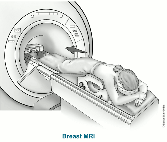
You’ll lie face down on a narrow, flat table with your arms above your head. Your breasts will hang down into an opening in the table so they can be scanned without being compressed. The technologist may use pillows to make you comfortable and help keep you from moving. The table then slides into a long, narrow tube.
The test is painless, but you have to lie still inside the narrow tube. You may be asked to hold your breath or keep very still during certain parts of the test. The machine may make loud thumping, clicking, and whirring noises, much like the sound of a washing machine, as the magnet switches on and off. Some facilities give you earplugs or headphones to help block noise out during testing.
When breast MRI is done to look for breast cancer, a contrast material called gadolinium is injected into a vein in the arm during the exam, which helps show any abnormal areas of breast tissue. (This is different from the contrast dye used in CT scans.) Let the technologist know if you have any allergies or have had problems before with any contrast or dye used in imaging tests.
It’s important to stay very still while the test is being done, which helps ensure the images will be of good quality.
Each set of images usually takes a few minutes, and the whole test usually takes about 30 to 45 minutes. After the test, you may be asked to wait while the pictures are checked to see if more are needed.
For a newer MRI technique, known as abbreviated breast MRI, fewer images are taken, so the scan takes less time (usually about 10 minutes).
How are breast MRI results reported?
Doctors use the same standard system to describe results of mammograms, breast ultrasound, and breast MRI. This system (called the Breast Imaging Reporting and Data System or BI-RADS) sorts the results into categories numbered 0 through 6.
By sorting the results into these categories, doctors can describe what they find on a breast MRI using the same words and terms. This makes communicating about these test results and following up after the tests much easier.
For more details on the BI-RADS categories, see Understanding Your Mammogram Report. While the categories are the same for each of these types of imaging tests, the recommended next steps after these tests might be different.
- Written by
- References
The American Cancer Society medical and editorial content team
Our team is made up of doctors and oncology certified nurses with deep knowledge of cancer care as well as editors and translators with extensive experience in medical writing.
American College of Radiology. ACR BI-RADS ATLAS – Breast MRI. Reporting System. 2013. Accessed at https://www.acr.org/-/media/ACR/Files/RADS/BI-RADS/MRI-Reporting.pdf on November 29, 2021.
Esserman LJ, Joe BN. Diagnostic evaluation of suspected breast cancer. UpToDate. 2021. Accessed at https://www.uptodate.com/contents/diagnostic-evaluation-of-suspected-breast-cancer on October 11, 2021.
Gupta D, Mendelson EB, Karst I. Nipple discharge: Current clinical and imaging evaluation. Am J
Roentgenol. 2021;216(2):330-339.
National Comprehensive Cancer Network. NCCN Clinical Practice Guidelines in Oncology. Breast Cancer. Version 8.2021. Accessed at https://www.nccn.org/professionals/physician_gls/pdf/breast.pdf on October 11, 2021.
Slanetz PJ. MRI of the breast and emerging technologies. UpToDate. 2021. Accessed at https://www.uptodate.com/contents/mri-of-the-breast-and-emerging-technologies on October 11, 2021.
Weinstein SP, Roth SO. Chapter 12: Imaging Analysis: Magnetic Resonance Imaging. In: Harris JR, Lippman ME, Morrow M, Osborne CK, eds. Diseases of the Breast. 5th ed. Philadelphia, Pa: Lippincott Williams & Wilkins; 2014.
Last Revised: January 14, 2022
American Cancer Society medical information is copyrighted material. For reprint requests, please see our Content Usage Policy.
American Cancer Society Emails
Sign up to stay up-to-date with news, valuable information, and ways to get involved with the American Cancer Society.

