Your gift is 100% tax deductible.
Mammograms
Mammograms are low-dose x-rays that can help find breast cancer. Learn all about mammograms, including their benefits and limitations, what their results might mean, and tips for getting one.
Why do I need mammograms?
Mammograms are used as a screening test for breast cancer in women without symptoms. They are also used for women who have symptoms that might be from cancer.
A mammogram can often find or detect breast cancer early, when it’s still small. Sometimes a mammogram can find breast cancer even before you feel a lump. When breast cancer is found early, treatment is more likely to be successful.
Types of mammograms
Mammograms are used in 2 main ways.
Screening mammograms are used to look for signs of breast cancer in women who don’t have any breast symptoms or problems. X-ray pictures of each breast are taken, typically from 2 different angles.
Diagnostic mammograms are used to look at a person’s breast if they have breast symptoms or if something unusual is seen on a screening mammogram. They might include extra views (images) of the breast that aren’t part of screening mammograms. Sometimes diagnostic mammograms are used for people who were treated for breast cancer in the past.
How do mammograms work?
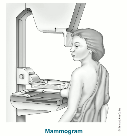
Mammograms are done with a special x-ray machine designed to look only at breast tissue.
During the mammogram, the breast is placed between 2 plates that compress or flatten the breast to spread the tissue apart. This gives the best quality picture and allows the least amount of radiation to be used. Pictures are taken from at least 2 angles to have the best chance of finding breast cancer early.
Mammograms can often show abnormal areas in the breast. They can’t tell for sure if an abnormal area is cancer, but they can help healthcare providers decide if more testing, such as a breast biopsy, is needed.
In the past, mammograms were typically printed on large sheets of film. Today, digital mammograms are much more common. Digital images are recorded and saved as files on a computer.
To learn more about how they are done, see Tips for Getting a Mammogram.
What are 3D mammograms?
Three-dimensional (3D) mammography is also known as breast tomosynthesis or digital breast tomosynthesis (DBT).
The experience of getting a 3D mammogram is much like that for a standard (2D) mammogram.
For a 3D mammogram, while each breast is compressed, the machine takes many images as it moves in a small arc around the breast. A computer puts the images together into a series of thin slices. This allows doctors to see the breast tissues more clearly in 3 dimensions.
A standard (2D) mammogram can be taken at the same time, or it can be reconstructed from the 3D mammogram images.
Potential benefits and limitations
Many studies have found that 3D mammography appears to lower the chance of being called back for follow-up testing after screening. It also appears to find some breast cancers better and be helpful in women with dense breasts.
However, 3D mammograms often cost more than 2D mammograms, and this added cost might not be covered by insurance.
Are mammograms safe?
While mammograms are generally considered safe, they do result in radiation exposure, some discomfort during the procedure, and sometimes extra tests that carry risks. The benefits of breast cancer screening outweigh any possible harm from the radiation exposure.
Mammograms and radiation
Modern machines use low radiation doses to get breast x-rays that are high in image quality.
Radiation can be measured in millisieverts (mSv). On average the total dose for a typical mammogram with 2 views of each breast is about 0.4 mSv. The radiation dose from 3D mammograms can range from slightly lower to slightly higher than that from standard 2D mammograms.
The dose of radiation used for a screening mammogram of both breasts is about the same amount of radiation a person would get from normal, everyday life over 1 to 2 months.
Mammograms during pregnancy
If there’s any chance you might be pregnant, let your healthcare provider and x-ray technologist know. Although the risk to the fetus is very small, and mammograms are generally thought to be safe during pregnancy, screening mammograms aren’t routinely done in pregnant people who aren't at increased risk for breast cancer.
Extra tests
Mammograms can also result in some people getting additional tests that don't result in a breast cancer diagnosis but that might still have their own risks.
Learn more in Limitations of Mammograms.
How are mammograms read?
A radiologist will look at your mammogram. A radiologist is a doctor who diagnoses diseases and injuries using imaging tests, such as x-rays.
Doctors use a standard system called the Breast Imaging Reporting and Data System (BI-RADS) to describe what they see on a mammogram.
Depending on where you have your mammogram done, the results might be ready right away, or a radiologist might review it later after you’ve left the testing center. Newer technologies might also be used to help radiologists find and evaluate suspicious areas on a mammogram.
Comparing new findings to your old mammograms
When possible, the doctor reading your mammogram will compare it to your old mammograms. This can help show if any findings are new, or if they were already there. Findings that haven’t changed from older mammograms aren’t likely to be cancer, which might mean you won't need further tests.
Understanding mammogram results
Mammogram results are compiled in a mammogram report. The radiologist reading your mammogram will look for different types of breast changes that could be signs of cancer.
Calcifications are tiny calcium deposits within the breast tissue. They look like small white spots on a mammogram. They might or might not be caused by cancer. There are 2 types of calcifications.
Macrocalcifications are larger calcium deposits that are most likely due to changes caused by aging blood vessels or skin, old injuries, or inflammation. They become more common after age 50. Macrocalcifications can be a variety of shapes and sizes, like rods or spheres. They are typically related to noncancerous conditions and don’t need further testing with a biopsy.
Microcalcifications are tiny specks of calcium in the breast. When seen on a mammogram, they are more of a concern than macrocalcifications, but they don’t always mean that cancer is present. The shape and layout of microcalcifications and whether they are near a mass help the radiologist judge how likely it is that the change is due to cancer.
If microcalcifications have a suspicious look or pattern, a biopsy will be recommended to check for cancer.
A mass is an area of abnormal breast tissue with a shape and edges that make it look different from the rest of the breast tissue on a mammogram. Masses can be many noncancerous things, but they might also be a sign of cancer.
When a mass is found, a radiologist will describe its shape, size, location, borders, and density on the mammogram. Certain features might make a mass more or less likely a cancer. A mass might be seen with or without calcifications.
Types of masses
Cysts are fluid-filled sacs. Simple cysts (fluid-filled sacs with thin walls) are not cancer and typically don’t need to be checked with a biopsy. If a mass is not a simple cyst (that is, if it’s at least partly solid, or if it has other concerning features), it’s of more concern. In that case, a biopsy might be needed to be sure it isn’t cancer.
Solid masses can be more concerning, but most solid breast masses are not cancer, such as fibroadenomas.
Determining the type of mass
A cyst and a solid mass can feel the same and look the same on a mammogram. A breast ultrasound is often done to determine whether a mass is a cyst because it is best at showing if a mass is solid or filled with fluid.
If it isn’t clear on an ultrasound if the mass is a cyst or a solid mass, a thin, hollow needle might be inserted into the mass during an ultrasound to try to remove (aspirate) the fluid from the area. If the mass goes away on the ultrasound as the fluid is removed, then it is most likely a cyst, and no further testing is needed.
If a mass is not a simple cyst, more imaging tests might be needed to decide if it could be cancer.
Some masses can be watched over time with regular mammograms or ultrasounds to see if they change, but others might need to be checked with a biopsy. The features of the mass can help the radiologist decide.
Asymmetries are areas of dense breast tissue that look different from the normal pattern and don’t have any calcifications. There are different types of asymmetries.
- Asymmetry: An area of dense breast tissue in one breast that looks different from the other breast, which can only be seen on 1 view of the mammogram.
- Focal asymmetry: A small area (less than one quadrant) of dense breast tissue that looks different from the other breast, which can be seen on 2 views of the mammogram.
- Developing asymmetry: An area of dense breast tissue that is new or larger when compared to prior mammograms and seen on 2 views of the mammogram. These changes are often followed up with a diagnostic mammogram and, if confirmed, lead to a biopsy.
- Global asymmetry: A large area (at least one quadrant) of dense breast tissue in one breast that looks different from the other breast, which can be seen on 2 views of the mammogram.
Most of the time, these findings do not mean you have breast cancer. But more imaging tests are often needed to make sure.
Radiologists use this term to describe when an area of the breast tissue appears distorted or pulled toward a certain point. Sometimes this might be due to how the breast was positioned during the mammogram. It might also be caused by a scar from a prior injury or procedure done on the breast. But it can also sometimes be a sign of breast cancer, so further imaging with diagnostic mammograms is typically needed to get a better look at this area.
Dense breasts have more fibrous and glandular tissue than fatty tissue. Breast density isn’t related to breast size or firmness.
About half of all women have dense breasts. Dense breast tissue is common and normal. However, women who have dense breasts have a slightly higher risk of breast cancer.
Dense breast tissue can also make it harder to see cancers on a mammogram. Normal fibrous and glandular tissue appears white on a mammogram which can hide other suspicious findings that also appear white. Still, experts don’t agree what other tests, if any, should be done along with mammograms in people with dense breasts who aren’t otherwise at higher risk for breast cancer.
Mammograms in special circumstances
If you have had breast cancer in the past, whether you need to keep getting mammograms might depend on the type of surgery you had.
If you have breast implants, you should get mammograms as recommended. But you might need to have extra pictures taken so the doctor can see as much breast tissue as possible.
- Written by
- References

Developed by the American Cancer Society medical and editorial content team with medical review and contribution by the American Society of Clinical Oncology (ASCO).
Aminololama-Shakeri S, Muhar R. Chapter 3: Mammographic Masses, Asymmetries, and Distortions. In: Joe BN, Lee AY, eds. Breast Imaging. 4th ed. Philadelphia, Pa: Elsevier; 2023.
Conant EF, Barlow WE, Herschorn SD, et al. Association of Digital Breast Tomosynthesis vs Digital Mammography With Cancer Detection and Recall Rates by Age and Breast Density. JAMA Oncol. 2019;5(5):635-642.
Durand MA, Friedewald SM, Plecha DM, et al. False-Negative Rates of Breast Cancer Screening with and without Digital Breast Tomosynthesis. Radiology. 2021;298(2):296-305.
Elmore JG, Lee CI. Screening for breast cancer: Evidence for effectiveness and harms. UpToDate. 2026. Accessed at https://www.uptodate.com/contents/screening-for-breast-cancer-evidence-for-effectiveness-and-harms on April 7, 2026.
Esserman LJ, Joe BN. Diagnostic evaluation of suspected breast cancer. UpToDate. 2026. Accessed at https://www.uptodate.com/contents/diagnostic-evaluation-of-suspected-breast-cancer on April 7, 2026.
Expert Panel on Breast Imaging:, diFlorio-Alexander RM, Slanetz PJ, et al. ACR Appropriateness Criteria® Breast Imaging of Pregnant and Lactating Women. J Am Coll Radiol. 2018;15(11S):S263-S275.
Greenwood HI. Chapter 2: Mammographic Analysis of Breast Calcifications. In: Joe BN, Lee AY, eds. Breast Imaging. 4th ed. Philadelphia, Pa: Elsevier; 2023.
Kruskal JB. Diagnostic imaging in pregnant and lactating patients. UpToDate. 2026. Accessed at https://www.uptodate.com/contents/diagnostic-imaging-in-pregnant-and-lactating-patients on April 7, 2026.
Lowry KP, Coley RY, Miglioretti DL, et al. Screening performance of digital breast tomosynthesis vs digital mammography in community practice by patient age, screening round, and breast density. JAMA Netw Open. 2020;3(7):e2011792.
Oeffinger KC, Fontham ET, Etzioni R, et al. Breast cancer screening for women at average risk: 2015 guideline update From the American Cancer Society. JAMA. 2015;314(15):1599-1614.
Pisano ED, Hendrick RE, Yaffe MJ, et al. Diagnostic accuracy of digital versus film mammography: exploratory analysis of selected population subgroups in DMIST. Radiology. 2008;246(2):376-383.
Pisano ED, Gatsonis C, Hendrick E, et al. Diagnostic performance of digital versus film mammography for breast-cancer screening. N Engl J Med. 2005;353(17):1773-1783.
Skaane P, Bandos AI, Niklason LT, et al. Digital Mammography versus Digital Mammography Plus Tomosynthesis in Breast Cancer Screening: The Oslo Tomosynthesis Screening Trial. Radiology. 2019;291(1):23-30.
Slanetz PJ, Lee CI. Breast imaging for cancer screening: Mammography and ultrasonography. UpToDate. 2026. Accessed at https://www.uptodate.com/contents/breast-imaging-for-cancer-screening-mammography-and-ultrasonography on April 7, 2026.
Weigel S, Heindel W, Decker T, et al. Digital Breast Tomosynthesis versus Digital Mammography for Detection of Early-Stage Cancers Stratified by Grade: A TOSYMA Subanalysis. Radiology. 2023;309(3):e231533.
Yaffe MJ, Mainprize JG. Risk of radiation-induced breast cancer from mammographic screening. Radiology. 2011;258(1):98-105.
Zackrisson S, Lång K, Rosso A, et al. One-view breast tomosynthesis versus two-view mammography in the Malmö Breast Tomosynthesis Screening Trial (MBTST): a prospective, population-based, diagnostic accuracy study. Lancet Oncol. 2018;19(11):1493-1503.
Last Revised: July 23, 2026
American Cancer Society medical information is copyrighted material. For reprint requests, please see our Content Usage Policy.
American Cancer Society Emails
Sign up to stay up-to-date with news, valuable information, and ways to get involved with the American Cancer Society.